

The Microbiological Landscape of the Microbiome
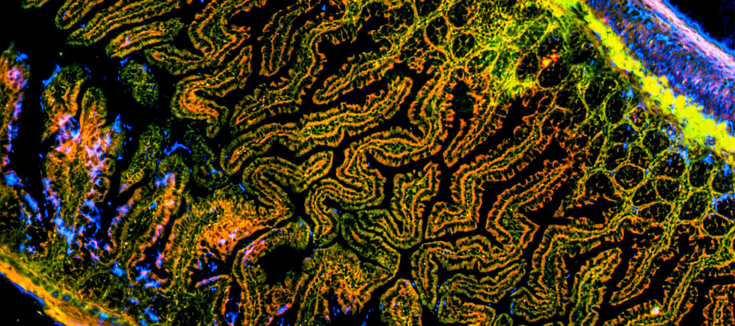
When people talk about the microbiome today, one term comes up almost automatically: sequencing. Lists of hundreds of taxa, relative frequencies with two decimal places. At first glance, this may seem impressive. But it raises a simple question: do we really understand what happens in the microbiome?
What Is Missing?
The sober answer is: only partially. Sequencing provides us with a list of names. But it does not tell us where these microorganisms are located in the system, with whom they occur together, whether they are active, whether they are organized in biofilms or occur in isolation. Stated frequencies convey a deceptive accuracy, although it is known that this level of detail cannot be achieved by sequencing.
Above all, one thing is missing: the picture.
The Telephone Directory
This is a problem for microbiome research and the development of probiotics and living biotherapeutics. After all, it is not just who is there, but how these microorganisms are organized as a group that is decisive. Whether a strain is only hidden in the sample as a DNA signal or is present as a living population makes a fundamental difference. Sequencing also represents DNA from dead cells and from freely circulating fragments. But the actual microbial reality remains invisible.
Relying solely on sequencing means analyzing the microbiome like a city using only the telephone directory. You know names and addresses, but not who meets where or which neighborhoods are lively. Only visualization shows which microorganisms actually perform the key functions and where problem areas are forming.
Really Understanding the Microbiome
This is exactly where the need for visualization begins. If you want to understand the microbiome, you need to see microorganisms directly in their habitat. Morphology, localization, associations with other populations, embedding in the matrix, density and distribution - all of this is only revealed through visualization. Only when bacteria, yeasts and archaea become visible in the true sense of the word can hypotheses from sequencing be verified or refuted.
VIT® gene probe technology, the industrialized further development of the FISH method, fluorescence in situ hybridization, can do just that. Specific, fluorescently labeled gene probes bind to the ribosomal RNA of living target organisms in the sample. These cells begin to glow under the microscope. This makes it possible to immediately recognize which microorganisms are actually alive, how many there are and where they are located. Without cultivation, without lengthy detours.
Direct Microbiology
At vermicon, we have been consistently developing this direct microbiology for almost three decades. Our VIT® gene probe technology and the services based on it enable the specific visualization and quantification of defined bacterial groups and species directly in complex samples. For microbiome projects, probiotics and living biotherapeutics, this means that we bring spatial structure, viability and absolute cell numbers into a field that is otherwise dominated by relative abundances and statistical estimates.
A preparation that appears stable in sequencing may turn out to be a cluster of a few surviving cells in a sea of foreign flora under the microscope. Conversely, visualization sometimes reveals robust, dense populations of a desired strain, while pure sequence analysis underestimates their significance. If you really want to control quality, you need both: the genetic information and the direct image of the system.
From Sequence Data to the Microbial Landscape
The visualization of microorganisms in the microbiome is therefore not a luxury, but a prerequisite for reliable results.
If you want to know what your microbiome, your probiotics or your living biotherapeutics look like in reality, we at vermicon will support you with VIT®-based visualization, specific quantification and sound advice. So that you don't just see names and sequences, but the actual microbial landscape you are working with.